Introduction
Cytogenetic analysis is a recommended test in newly diagnosed AML but requires accurate interpretation of the International System for Human Cytogenomic Nomenclature (ISCN). Keeping up to date across recent published classifications - WHO, the European Leukaemia Net (ELN) and International Consensus Classification (ICC) - is critical for accurate prognostic assessment, but can be challenging for clinicians and laboratory scientists. Furthermore, eligibility to access state-funded treatments in the UK is dictated by certain cytogenetic results. We previously developed a webapp that integrates cytogenetic, molecular and clinical features to aid accurate classification, prognostication and provide treatment recommendations in AML (Coats et al, BJH 2021). To further improve the webapp we have developed a cytogenetic classifier to automatically interpret reports written in ISCN format.
In this study we aimed to validate the automated cytogenetic classifier (ACC) in identifying abnormalities that feature in the new WHO, ELN and ICC classifications.
Methodology
Software for the ACC was written in Python to:
1. count the number and type of karyotypic abnormalities (subdivided into number of monosomies, complete chromosome gains and structural changes)
2. identify presence of clinically relevant, named, abnormalities that feature in international classifications (categorised as an individual abnormalityg. t(9;11)(p21.3;q23.3), or a generic abnormality e.g. any translocation involving 11q23.3).
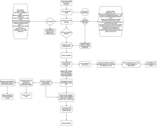
Cytogenetic reports of 50 consecutive AML cases with abnormal karyotypes analysed by Bristol Haematological Oncology Diagnostic Service (BHODS) were selected (training cohort). Each report was uploaded to the ACC and its output independently checked for accuracy. Following review of the software's performance, adjustments to the ACC were made. The updated ACC (Figure 1) analysed 98 additional reports from two other laboratories, UCLH and OUH (validation cohort).
Results
The ACC analysed 49/50 cytogenetic reports from the BHODS laboratory. One report could not be read by the ACC. The correct quantity of abnormalities was identified in 49/49 cases analysed. Where relevant named abnormalities occurred, these were detected in 79/80 instances. One case of acute promyelocytic leukaemia containing an idic(17p) was not identified as containing an abnormal 17p. Additionally, in one report with a complex karyotype, a monosomy 13 was erroneously detected by the ACC.
Adjustments were made to the ACC such that it could analyse all 50 cases with 100% accuracy. The updated ACC then analysed 98 cases from the validation cohort. The correct quantity of abnormalities was detected in 97/ 98 cases. In one case with a highly complex karyotype containing a tetraploid clone, the ACC incorrectly counted 48, as opposed to 45, abnormalities. Clinically relevant, named, abnormalities were correctly identified in 178/179 instances. In one case, containing a 4-way translocation, the ACC did not identify the t(9;11) abnormality.
Discussion
Here, we show that the updated ACC is accurate in identifying the number and type of karyotypic abnormalities relevant to AML from different laboratories. Reduced accuracy in counting the number of abnormalities in highly complex karyotypes (i.e. >40 abnormalities) is not likely to impact clinical interpretation. Further validation work is needed to analyse 3 and 4-way translocations. User warnings have been added to the ACC for karyotypes containing multiple-translocations and tetraploid/triploid clones to improve clinical safety.
The ACC can support clinical decision-making by streamlining cytogenetic interpretation from across multiple clinical classifications. It can be used as a stand-alone programme or integrated into more sophisticated decision support tools. The ACC has the potential to be updated to interpret cytogenetic reports from other classifications or haematological malignancies.
The ACC is available as a webapp (Streamlit (ts482-cytogenetics-calculator-extractorstreamlit-w7dq9p.streamlit.app).
Figure 1. Flowchart of the methodology of the updated automated cytogenetic classifier (ACC).
Disclosures
No relevant conflicts of interest to declare.


This feature is available to Subscribers Only
Sign In or Create an Account Close Modal